Cellularis® Discovery - EarlySight
CELLULARIS® Discovery - See the Invisible
The disruptive ophthalmic camera from EarlySight, delivers cellular level images of various structures of the retina and optic disc to investigate the eye anatomy and the pathological processes in a completely new perspective.
Lateral illumination
Revolutionary patented non-contact transscleral flood illumination creates contrast to hidden cells and biological structures affected by eye diseases, such as retinal pigment epithelium cells.
The “non-invasive biopsy”
A revamped and easy-to-use adaptive optics technology brings imaging resolution to a completely new level. Look at single cells composing the tissue, as if under a microscope, observe pathological processes in details.
Fundus overview
Optimize your examination workflow by targeting immediately the area of interest. Cellularis offers a widefield fundus overview image simultaneously with the cellular level image to easily locate the imaged area within the eye.
Patient-centric design with efficient user experience
Fast and comfortable image acquisition for the patient to easily locate the imaged area within the eye. Ergonomic and easy-to-use instrument to facilitate eye examinations.
Share


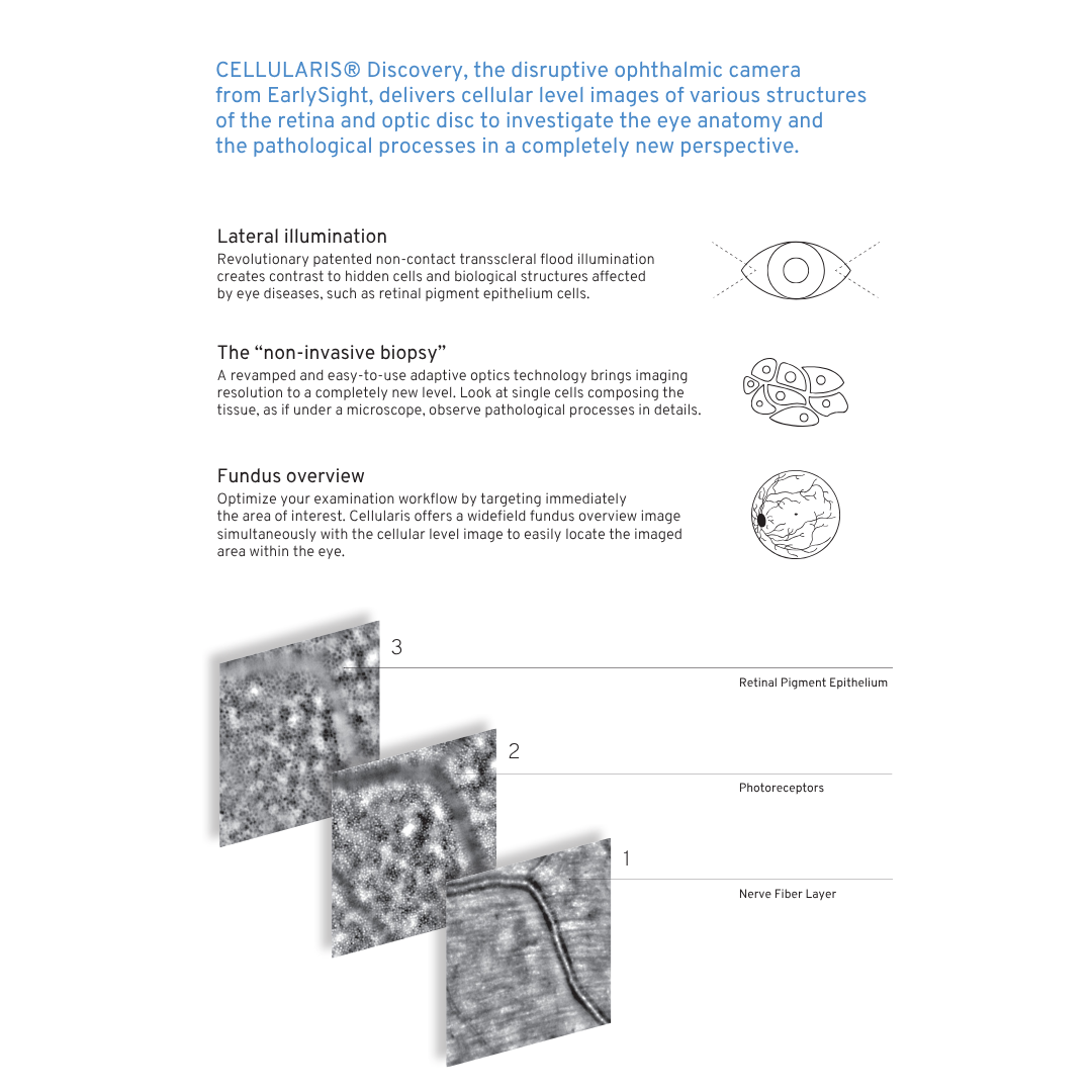
Look at Tissue Details
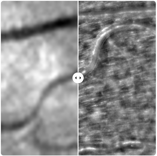
As opposed to standard instruments, our optical technology allows the observation of microscopic structures in the eye. The ideal tool to catch the first impacts of tissue degenerative processes.
Benefits
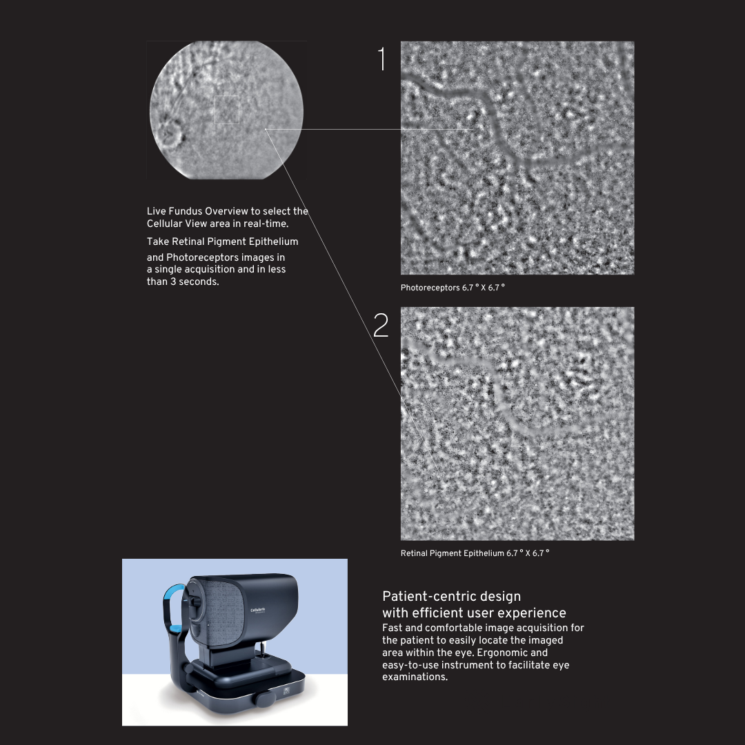
Cellularis Discovery provides images that are 10 times more detailed than the standard instruments used in hospitals. Its spectacular precision is achieved while providing a great user experience. Indeed, a quick acquisition time together with manual joystick operation enable simple and familiar examination process for both patient and operator. Moreover, infrared lateral and transpupil illumination is very comfortable for the patient.